Protect your vision.
Bringing the most advanced technology to our patients, we offer optomap® ultra-wide digital retinal imaging as part of your comprehensive eye exam today.
The optomap ultra-wide digital retinal imaging system helps you and your eye care professional make informed decisions about your eye health and overall well-being. Combining your eye care professional’s expertise and optomap technology,optomap brings your eye exam to life.
PIONEERING TECHNOLOGY

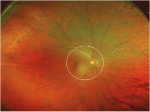
optomap ultra-wide digital retinal image of a healthy eye with the traditional view indicated by the circle.

This device is intended for operation by trained eye care professionals only. Your eye care professional will advise you whether the optomapexam is suitable for you. Use only as directed.
Early Detection is Vital
What is your retina?
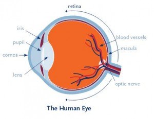
The retina is a delicate lining at the back of the eye similar to film in a camera. Light strikes the retina through the lens in your eye and produces a picture which is then sent to the brain, enabling you to see.
Why is a healthy retina important?
An unhealthy retina cannot send clear signals to your brain which can result in impaired vision or blindness. Many retinal conditions and other diseases can be treated successfully with early detection.

Without a comprehensive eye exam, you may not be aware of a potential problem. You may see clearly, and because the retina has no nerve endings, you may not feel any pain, a symptom which may otherwise prompt you to see your eye care professional.
What can happen to the retina?
Your retina is the only place in the body where blood vessels can be seen directly. This means, in addition to eye conditions, signs of some systemic diseases can be seen in the retina. Early detection is important so treatments can be administered.
How does your eye care professional normally examine the retina?
Examining the retina is challenging. Your eye care professional looks through your pupil to examine the back of your eye. Traditional viewing methods can be effective, but difficult to perform and are carried out manually without any digital record.
How does the optomap help?
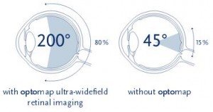
The optomap ultra-wide digital retinal imaging system captures more than 80% of your retina in one panoramic image. Traditional methods typically reveal only 10-15% of your retina at one time.
Seeing most of the retina at once allows your eye care professional more time to review your images and educate you about your eye health. Numerous studies have demonstrated the power of optomap as a diagnostic tool1.
Do all eye care professionals have an optomap ultra-wide digital retinal imaging system?
optomap is a standard of care for evaluating eye health in this office and millions of people worldwide have benefited from optomap.
How often should I have an opto map?
Your eye care professional will advise you based on your individual circumstances,but the general recommendation is that you have an opto map every time you have an eye exam. This will ensure you have a digital record of your retinal health on file which can be compared for changes over time.
Should my children have an optomap too?
Many vision problems begin at an early age, so it’s important for children to receive proper eye care from the time they are infants.
Will I need to be dilated and does it hurt?
An opto map takes only seconds to perform, is not painful, and typically does not require dilation. However, your eye care professional may decide dilation is still needed.
More than 37 million optomaps have been performed worldwide since 2000.
An important method for evaluating eye health.
How was optomap invented?
“In 1990 my five year old son Leif Anderson went blind in one eye because a retinal problem was detected too late for treatment. Although he was having regular eye exams, conventional tests were uncomfortable, especially for a small child. I sought to find a way to make retinal examinations easier. Leif, now a young man, has adjusted beautifully and we are thankful to, hopefully, help other families avoid vision loss.” —Douglas Anderson, Optos founder


